The specialists at ProVascularMD have compiled an extensive gallery of arterial ulcer pictures to help you better understand what’s going on with your legs. We provide more explanation on the different visual features of peripheral artery disease in more detail below the gallery. You can also check out our galleries on
venous insufficiency pictures and
venous ulcer pictures to learn more about other vascular conditions that affect the legs.
Arterial Ulcer Treatment in Los Angeles
See a Vascular Ulcer Specialist
Arterial Ulcer Visual Features
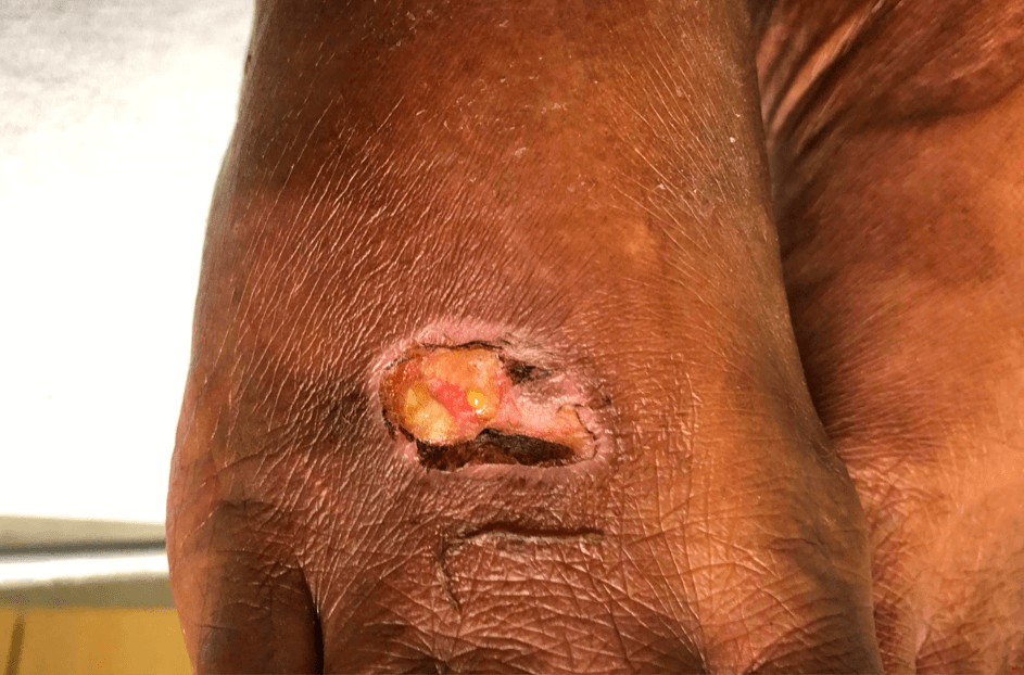
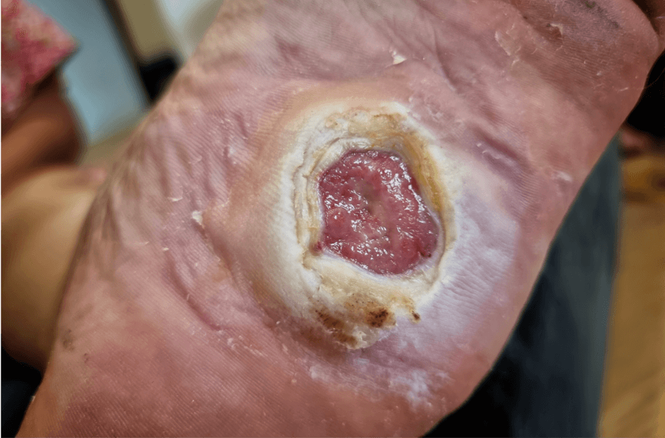
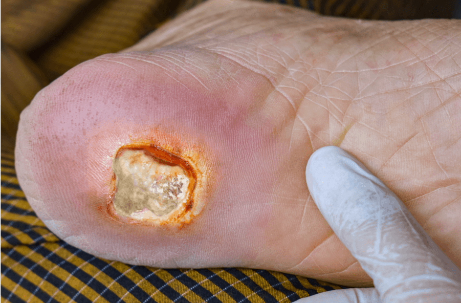
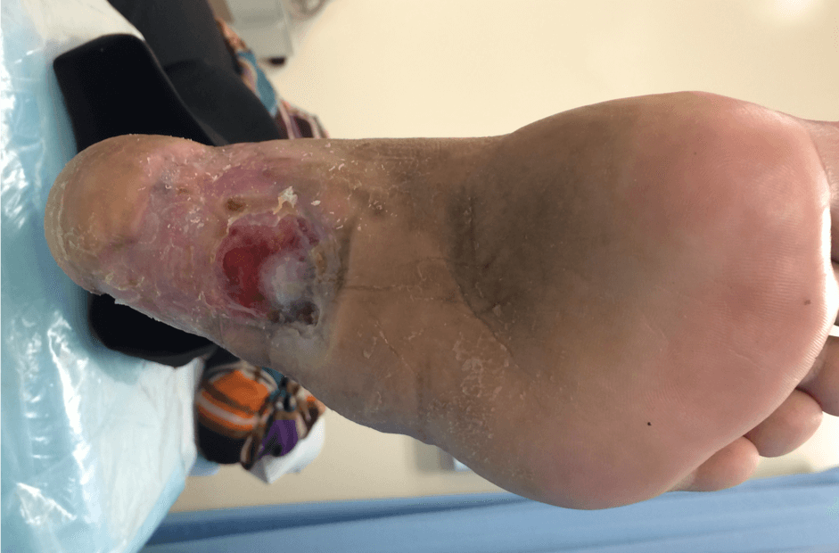
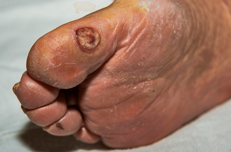
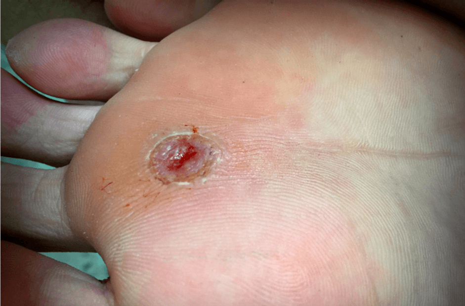
Arterial ulcers typically present as sharply demarcated, round or oval lesions with steep, cliff-like edges. The borders appear clean, as if a portion of the skin has been removed neatly. This is due to the lack of blood flow, which prevents the wound from healing properly or forming granulation tissue, giving the ulcer its distinct, “punched-out” look. These ulcers are often found on pressure points, such as the toes, feet, or ankles.
Shallow or Deep Ulceration
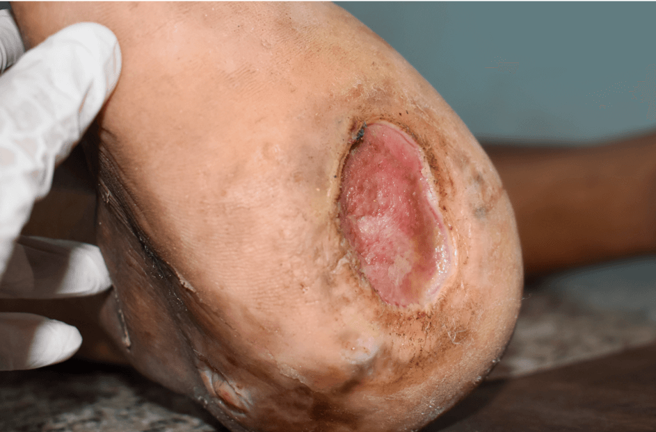
Arterial ulcers often start as superficial, shallow wounds but can extend deep into the skin and underlying tissues. In advanced cases, they may penetrate through the dermis and subcutaneous layers, exposing structures like tendons, muscles, or even bone. The depth is typically indicative of the severity of arterial insufficiency, and deeper ulcers are more challenging to heal due to compromised blood flow.
The base of the ulcer may appear pale, gray, or yellowish because of poor oxygenation and reduced blood flow, resulting in minimal granulation tissue formation. This is in stark contrast to venous ulcers, which usually have a red or pink wound bed. The lack of redness signifies that the healing process is impaired, and the tissue is not regenerating as it should.
Arterial ulcers tend to be dry and produce little to no drainage (exudate). The reduced exudate is a result of the decreased blood supply, which limits the production of inflammatory fluids and inhibits the body’s natural healing process. This dryness is one of the distinguishing features from venous ulcers, which often produce more exudate due to excessive pressure and fluid buildup.
Shiny, Thin, Hairless Skin
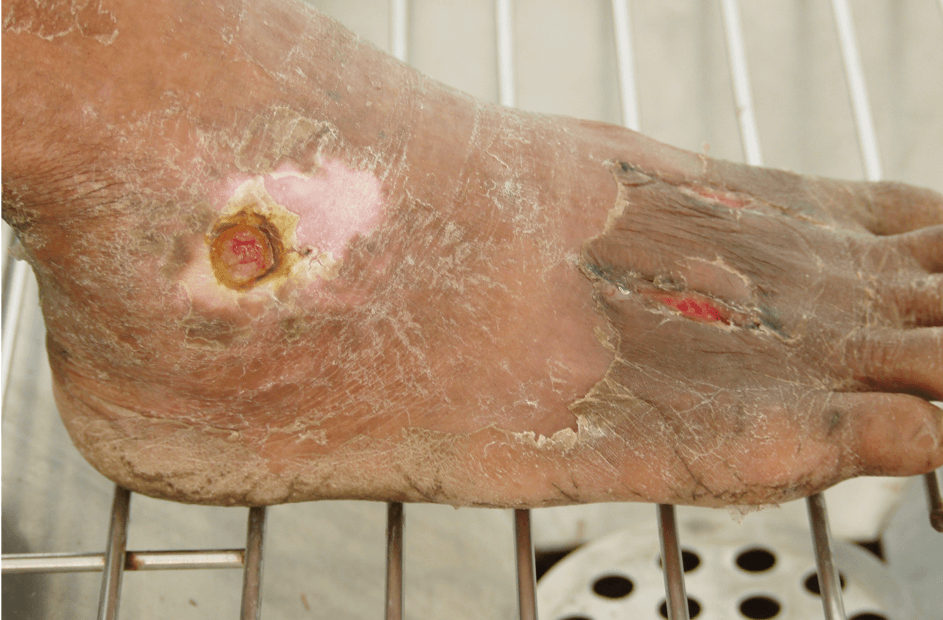
The skin around an arterial ulcer often looks shiny, thin, and may be hairless due to prolonged ischemia (reduced blood flow). The lack of hair growth is a result of poor circulation, which starves the hair follicles of nutrients. Over time, the skin becomes atrophic, appearing taut, dry, and almost translucent. This is a classic sign of peripheral arterial disease (PAD), which is often associated with arterial ulcers.
When the leg is elevated (e.g., during a physical exam), the skin may turn pale due to gravity’s effect on blood flow. In patients with arterial insufficiency, the already limited blood supply struggles to reach the extremities when elevated, causing a blanching or whitening of the skin. This is a classic sign of poor arterial perfusion and can be tested by raising the leg above heart level for a few minutes.
Redness or Rubor When Dependent
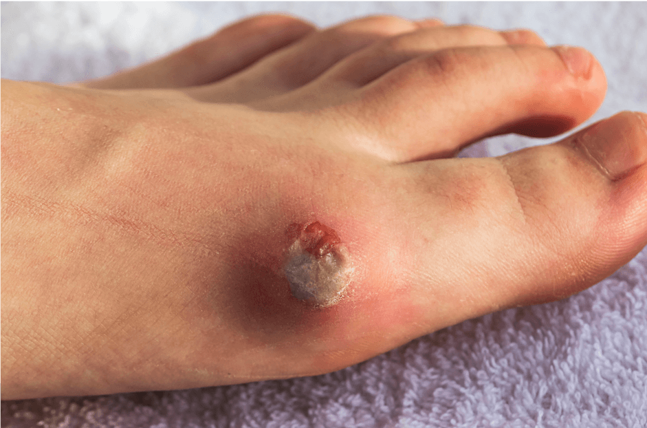
When the leg is lowered back down, it may turn a dark red or purplish color (dependent rubor) due to blood pooling in the extremities. This occurs because the arteries are unable to constrict properly in response to gravity. The blood collects in the tissue instead of flowing efficiently, leading to the discoloration. This color change is a key clinical indicator of severe arterial disease.
Surrounding Tissue is Cool
The skin surrounding the ulcer often feels cool or cold to the touch, reflecting the diminished blood circulation to the affected area. This coolness is particularly noticeable when compared to the temperature of healthy tissue in other parts of the body. This sign of poor perfusion is key in diagnosing arterial ulcers, as the coolness indicates the lack of warm, oxygen-rich blood.
Palpation of the pulses in the foot (such as the dorsalis pedis or posterior tibial pulse) is often weak or completely absent in patients with arterial ulcers. This absence of pulses is a strong indicator of peripheral arterial disease (PAD) and reflects the severe restriction of blood flow to the lower extremities. Doppler ultrasound is often used to assess the degree of arterial blockage.
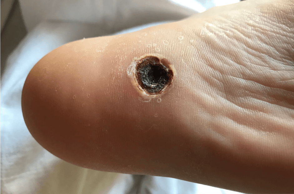
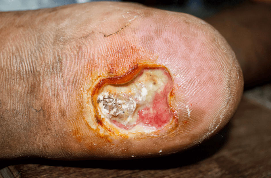
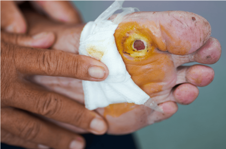
The presence of necrotic (dead) tissue is common in arterial ulcers. The necrotic tissue can range from yellowish slough (dead cells) to dry, black eschar (a thick, leathery crust of dead tissue). The eschar forms as the tissue dies from a lack of blood and oxygen. In some cases, the eschar is hard and adheres firmly to the wound bed, preventing the wound from healing until it is debrided (removed).
In severe cases, arterial ulcers may progress to gangrene, a condition where the tissue turns black and necrotic. This is a result of complete tissue death due to the total lack of blood supply. Gangrene can affect the ulcer itself or the surrounding tissues, leading to potential amputation if left untreated. It presents as black, leathery, and often foul-smelling tissue.
Arterial Ulcer Treatment in Los Angeles
See a Vascular Ulcer Specialist
Lower Extremity Arterial Disease
Learn what it is, how to spot symptoms, and your treatment options in this quick overview.
Peripheral Artery Disease (PAD) Screening
Learn why peripheral artery disease screening is essential in the battle against amputation.
Peripheral Arterial Disease Treatment
A brief 101 on the treatment options available for arterial disease.
What Are Arterial Ulcers?
Signs and symptoms of arterial ulcers, and how to treat them.
Vascular Specialist in Los Angeles
Learn more about Los Angeles Vascular Specialist Dr. Michael Lalezarian.