
Chronic Venous InsufficiencyDowney, CA
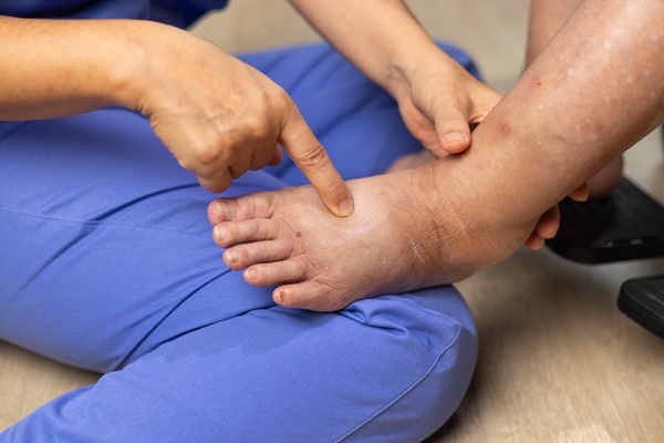
Chronic venous insufficiency is a condition that develops when the veins in the legs have difficulty pushing blood back toward the heart. Instead of moving upward as intended, blood begins to pool in the lower legs, creating pressure inside the veins and surrounding tissues. Over time, this pressure can lead to swelling, aching, heaviness, visible changes in veins, and skin damage that interfere with daily comfort and mobility.
Fortunately, ProVascularMD provides advanced, image-guided care to identify the source of venous disease and offer minimally invasive treatment options that support lasting relief. Our team understands that chronic venous insufficiency can affect both quality of life and long-term leg health, which is why we focus on timely evaluation and personalized care. If you live in or around the Downey area, we can help.
What Causes Chronic Venous Insufficiency?
Chronic venous insufficiency usually occurs when the valves inside the leg veins become weak or damaged. While these tiny valves normally keep blood moving upward in the right direction, blood can flow backward and pool in the lower extremities when they fail to close properly. This backward flow creates excess pressure inside the veins and gradually stretches them, worsening the problem over time.
Several risk factors can increase the likelihood of developing this condition. Various factors that can place excess strain on the venous system include:
- A history of blood clots
- Prolonged standing or sitting
- Pregnancy
- Excess weight
- Family history of vein disease
Age also plays a role, as the veins and their valves may weaken over time, making it harder for the legs to maintain healthy circulation.
As pressure builds inside the veins, the surrounding tissues begin to show the effects of poor circulation. Swelling may become more persistent by the end of the day, and the skin can become irritated, discolored, or fragile. Without treatment, chronic venous insufficiency may continue to progress and increase the risk of skin breakdown or venous ulcers near the ankles.
“Chronic venous insufficiency usually occurs when the valves inside the leg veins become weak or damaged.”
The Role of a Vascular and Interventional Radiologist in Treating Chronic Venous Insufficiency
A vascular and interventional radiologist is a specialist who uses advanced imaging technology to diagnose and treat circulation problems from inside the blood vessels. Instead of relying on large incisions, these specialists perform many vein treatments through a tiny puncture in the skin. This approach allows for precision, comfort, and shorter recovery times for many patients dealing with chronic venous insufficiency.
By using real-time imaging, a vascular and interventional radiologist can identify which veins are leaking, where blood is pooling, and how severely the venous system has been affected. Once we map out the problem veins, treatment can target the underlying cause of the pressure rather than only managing the symptoms. This focused approach helps improve blood flow, reduce swelling, and protect the skin from further damage.
“By using real-time imaging, a vascular and interventional radiologist can identify which veins are leaking, where blood is pooling, and how severely the venous system has been affected.”
What to Expect During a Consultation
The consultation begins with a detailed discussion about symptoms, daily discomfort, and any visible changes in the legs. We may ask whether swelling worsens throughout the day, if the legs feel heavy or tired, and whether there has been itching, discoloration, or skin breakdown near the ankles. These details help us understand how venous disease is affecting a patient’s comfort and function.
We will review the factors above and other signs, such as swelling or bulging veins, during a physical exam to look for signs of underlying venous reflux. In most cases, we conduct an ultrasound study to observe blood flow through the veins in real time and determine whether the valves are allowing blood to flow backward.
This visit also provides an opportunity to explain the diagnosis and outline possible treatment options. Patients can ask questions about recovery, symptom relief, and what to expect from minimally invasive care. The goal is to provide a clear path forward so that each person feels informed and prepared before treatment begins.
“In most cases, we conduct an ultrasound study to observe blood flow through the veins in real time and determine whether the valves are allowing blood to flow backward.”
What is Chronic Venous Insufficiency Treatment?
Chronic venous insufficiency treatment focuses on improving blood flow in the leg veins and reducing the pressure that causes swelling, pain, and skin damage. The right treatment depends on which veins are affected and how severe the symptoms have become. In many cases, minimally invasive procedures can treat the source of the problem without the need for traditional surgery.
One common treatment is venous ablation, which closes off a damaged vein so blood can reroute through healthier vessels. Sclerotherapy may also help treat certain abnormal veins by injecting a solution that causes them to collapse and fade. In some situations, we may recommend additional image-guided vein procedures to relieve pressure and improve overall venous circulation in the legs.
Treatment may also include compression strategies and recommendations that support healthy circulation between visits. Addressing the faulty veins early can reduce symptoms and help prevent the skin from becoming increasingly fragile or ulcerated. When paired with ongoing monitoring, minimally invasive vein treatment can lead to meaningful improvements in both comfort and long-term leg health.
“Chronic venous insufficiency treatment focuses on improving blood flow in the leg veins and reducing the pressure that causes swelling, pain, and skin damage.”
Benefits of Treating Chronic Venous Insufficiency
One of the biggest benefits of treatment is relief from the swelling, heaviness, and aching that often interfere with daily activity. When blood stops pooling in the lower legs, many patients notice that standing and walking feel easier and less exhausting. Reducing this pressure can also improve comfort at the end of the day, when symptoms often feel most intense.
Treatment also helps protect the skin and soft tissues from long-term damage. Chronic venous insufficiency can cause discoloration, irritation, thickening of the skin, and ulcers that are difficult to heal if the underlying pressure remains untreated. Improving venous circulation lowers the risk of these complications and supports healthier tissue over time.
Another important benefit is the opportunity to improve the quality of life without major surgery. Minimally invasive vein treatments often involve less downtime and faster recovery than traditional procedures. This allows patients to pursue meaningful symptom relief while returning to normal routines more quickly.
“One of the biggest benefits of treatment is relief from the swelling, heaviness, and aching that often interfere with daily activity.”
Questions Answered on This Page
Q. What causes chronic venous insufficiency
Q. What happens during a chronic venous insufficiency consultation?
Q. What treatments are available for chronic venous insufficiency?
Q. What are the benefits of treating chronic venous insufficiency?
Frequently Asked Questions
Q. What are the symptoms of chronic venous insufficiency?
A. Common symptoms include swelling in the lower legs, aching, heaviness, fatigue, and discomfort that worsens after long periods of standing or sitting. Some patients also notice varicose veins, itching, or a burning sensation in the legs. As the condition progresses, the skin may become discolored, thickened, or more prone to sores near the ankles.
Q. Is chronic venous insufficiency serious?
A. Chronic venous insufficiency can become serious when it is left untreated for a long period. Ongoing pressure in the veins may lead to worsening swelling, skin breakdown, and venous ulcers that are difficult to heal. Early evaluation helps reduce the risk of these complications and supports better long-term leg health.
Q. How is chronic venous insufficiency diagnosed?
A. Diagnosis usually begins with a review of symptoms and a physical examination of the legs. An ultrasound is commonly used to evaluate blood flow inside the veins and determine whether the valves are allowing blood to move backward. This test helps identify which veins are causing the problem and guides treatment planning.
Q. Can chronic venous insufficiency be treated without surgery?
A. Yes, many patients can be treated with minimally invasive procedures instead of traditional surgery. Options such as venous ablation and sclerotherapy can close or redirect faulty veins through a small puncture in the skin. These treatments often involve less downtime and allow patients to return to daily activities more quickly.
Q. When should a person seek a vascular evaluation for chronic venous insufficiency?
A. A vascular evaluation is important when swelling, aching, heaviness, skin changes, or visible veins continue to worsen or interfere with daily life. It is especially important to seek care when the skin near the ankles becomes discolored, irritated, or begins to break down. Early assessment can identify the cause of venous disease and help prevent more serious complications.
Call Us Today
Persistent leg swelling, aching, heaviness, or skin changes may be signs of chronic venous insufficiency that patients should not ignore. Early treatment can improve comfort, reduce pressure in the veins, and help protect the skin from more serious complications. Therefore, if you are experiencing symptoms of venous disease, contact ProVascularMD at (310) 341-4867. We provide specialized vascular care for patients in and around the Downey area who need answers and effective treatment for chronic vein problems.
Related Posts

What Can An Interventional Radiologist Treat?
A vascular and interventional radiologist treats a wide variety of medical conditions by using high-tech imaging to guide tools through tiny incisions in the skin. With this type of care, there is no need to make large surgical cuts. This specialty changes the way people experience medical care by offering solutions that once required hours…
Common Causes Of Leg Swelling
Leg swelling is a common symptom that can range from mild to severe and has many causes. Sometimes, the cause is temporary and of mild to moderate concern, but leg swelling may point to a more serious health condition in other cases. Learning more about leg swelling helps people know when to seek help from…

Find Relief From Knee Pain With Genicular Artery Embolization
Genicular artery embolization is a minimally invasive option for chronic knee pain linked to osteoarthritis, particularly when conservative treatments no longer provide enough relief. It is common to want improved walking comfort without moving straight to joint replacement. This procedure makes it possible for many patients. A vascular and interventional radiologist can perform this procedure…

What To Know About Prostate Artery Embolization
Prostate artery embolization offers a modern, gentle solution for men who live with the daily frustrations of an enlarged prostate. This specialized procedure provides relief without the need for traditional surgery or the removal of tissue. Men who seek an alternative to long-term medication or invasive operations often find that visiting with a vascular and…

Welcome To ProVascularMD
At ProVascularMD, we encourage patients to take control of their health and educate themselves on their condition as well as the treatment options we offer. We regularly publish blog posts on our site to provide patients with the tools they need to manage their condition and improve their health in various ways. Our blogs serve…