
Painful NeuropathyDowney, CA
Painful neuropathy can cause burning, tingling, stabbing sensations that cause ongoing discomfort. This condition often develops when nerves become irritated or damaged, leading to symptoms in the feet, legs, hands, or arms. While neuropathy is commonly linked to diabetes, it can also be the result of circulation issues, chronic inflammation, past injuries, and other medical conditions. A vascular and interventional radiologist can help determine whether blood flow problems are contributing to symptoms and provide treatment for better health and comfort.
At ProVascularMD, our team takes a personalized approach to managing painful neuropathy. We can help patients find clarity and a path forward, even when symptoms overlap with other vascular concerns. Call our Downey office at (310) 341-4867 to schedule an appointment today.
What Is Painful Neuropathy?
Painful neuropathy is nerve-related pain that occurs when peripheral nerves, which carry signals between the brain, spinal cord, and body, become damaged or dysfunctional. Symptoms include burning, pins-and-needles sensations, numbness with pain, or shooting pain. These symptoms tend to worsen at night or flare up after activity. Neuropathy can also reduce protective sensation, which raises the risk of unnoticed injuries, especially in the feet.
It is recommended to get a vascular evaluation when symptoms such as cold feet, cramping when walking, skin discoloration, slow-healing sores, or ulcers appear. These signs can suggest there is reduced blood flow or venous congestion, which can worsen nerve irritation and tissue sensitivity.
“Symptoms include burning, pins-and-needles sensations, numbness with pain, or shooting pain.”
How Different Causes of Painful Neuropathy Influence Symptoms
Common causes of painful neuropathy include:
- Diabetes and long-term elevated blood sugar
- Peripheral arterial disease (PAD) and reduced circulation to the legs and feet
- Chronic venous insufficiency and persistent leg swelling
- Vitamin deficiencies, thyroid disorders, or medication side effects
- Past injuries, nerve compression, or chronic inflammation
Neuropathy can present in different patterns due to the root cause. Diabetic neuropathy follows a stocking pattern, beginning in the toes and traveling up the leg. However, those with nerve compression tend to report more localized pain in a specific nerve region. Since multiple conditions can cause neuropathic pain, an evaluation by a vascular and interventional radiologist is often an essential step in treatment planning.
“Neuropathy can present in different patterns based on its cause.”
Diagnosis and Testing
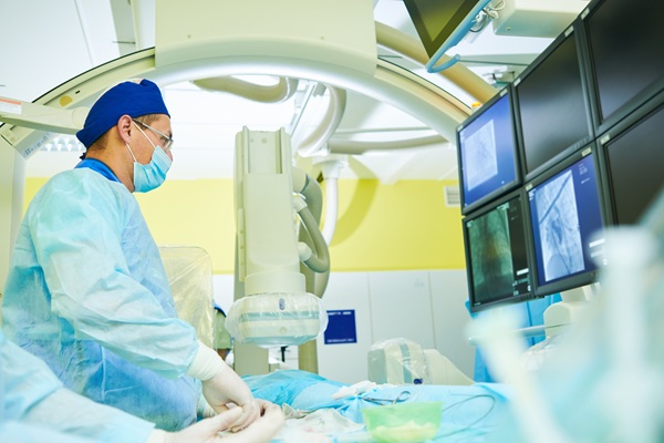
A vascular and interventional radiologist can test for neuropathy through a step-by-step process that reviews symptoms, risk factors, and circulation. This begins with a detailed history and physical exam. The goal is to clarify how symptoms present themselves, where pain is located, what triggers flare-ups, and whether any other vascular concerns are present.
Next, the provider will likely order non-invasive vascular testing to assess blood flow. Depending on symptoms, this may include ankle-brachial index (ABI) testing, a Doppler ultrasound, or other tests to look for conditions like peripheral arterial disease or chronic venous insufficiency. Imaging can help map arteries or veins and identify narrowing, blockages, or reflux that could be contributing to symptoms.
“The goal is to clarify how symptoms present, where pain is located, what triggers flare-ups, and whether any other vascular concerns are present.”
Vascular Care for Painful Neuropathy
When circulation problems are a root cause of a patient’s neuropathy, treatment may include one or more of the following:
- Arterial testing and treatment. Minimally invasive options such as angioplasty, stenting, or other arterial revascularization techniques based on blockage location and severity.
- Vein treatment options. For chronic venous insufficiency, varicose veins, or leg swelling, which may involve reducing pressure within the veins.
- Wound-focused care. When vascular ulcers or slow-healing sores are present, this approach uses monitoring designed to prevent complications and protect tissue health.
By improving blood flow, vascular care can support healthier tissue function, reduce pain triggers, and lower the risk of ulcers in patients with painful neuropathy. Moreover, when swelling decreases and circulation improves, mobility and daily comfort follow. Even when circulation is not the main cause, identifying that early helps streamline referrals and move patients toward the most effective treatment option.
“By improving blood flow, vascular care can support healthier tissue function, reduce pain triggers, and lower the risk of ulcers in patients with painful neuropathy”
The Importance of Early Collaborative Care
Painful neuropathy often improves most successfully when evaluation and care start early. This is particularly the case if more than one factor contributes to symptoms. A coordinated plan helps identify the root cause, reduce delays, and connect patients with the right support from the beginning.
For example, a vascular and interventional radiologist may collaborate with primary care providers, endocrinologists, podiatrists, and wound care teams, especially when diabetic neuropathy treatment and ulcer prevention are part of the picture. Not only can this expand treatment options, but it can also improve safety and support more predictable, long-term symptom relief.
“A coordinated plan helps identify the root cause, reduce delays, and connect patients with the right support from the beginning.”
What is a Spinal Cord Stimulator?
In many ways, your spine is the central nervous highway of your body. It is responsible for carrying all manner of neurological signals from your body to your brain, and from your brain to your body. Painful sensations are communicated through the spine, including pain caused by diabetic neuropathy. A spinal cord stimulator is a medical device that delivers electrical energy to the spine, therefore interrupting pain signals before they reach the brain.
When you receive a spinal cord stimulator for diabetic nephropathy, electrical stimulation is delivered to your spine by way of leads that are connected to an implantable pulse generator (an IPG) by small wires. Leads are specially designed electrical contacts that are placed near the spine. Electrical signals are sent to the leads by the IPG, which is a battery-powered micro-electronic device that can be programmed to deliver specific patterns of electrical stimulation. The IPG is controlled by an external remote control, which can be used to modify stimulation patterns, intensity, and other settings.
At a glance, a spinal cord stimulator device looks quite similar to a cardiac pacemaker, but a spinal cord stimulator has special features that are designed specifically for the spine and for pain therapy.
How Does Spinal Cord Stimulation Work in Diabetic Neuropathy?
Diabetic neuropathy is a condition in which nerves in the arms, hands, legs, feet, and other regions of the body are damaged by chronic, uncontrolled diabetes. Pain signals originate in the peripheral extremities and are communicated to the spine through specialized nerve fibers called A beta, A delta, and C fibers. From the spine, the signals are integrated and sent to the brain where they are processed and perceived. Damage to peripheral nerves can cause severe pain signaling even when there is no identifiable source of pain.
A spinal cord stimulator works by creating an electric field near the nerves along the spine. This electric field interferes with the electrical activity of the nerves that are involved with pain signaling to the brain. When the spinal cord stimulator is active, painful sensations are replaced with a mild tingling sensation.
An important advantage of a spinal cord stimulator is that it can be personalized to your pain patterns and sensory preferences. With an external remote control, the electrical energy can be targeted towards specific parts of your spine, delivered in different patterns, amplified, or reduced with the press of a few buttons.
What to Expect from Spinal Cord Stimulator Surgery
Unlike most medical procedures, spinal cord stimulator surgery requires two separate procedures. The first is a trial procedure, which is designed to help you decide whether or not the spinal cord stimulator provides you with pain relief. The second is the implant procedure, which only takes place if you experienced pain relief during the trial.
Spinal Cord Stimulator Trial
The spinal cord stimulator trial can be thought of as a test drive of the implantable device. Not every diabetic neuropathy patient finds pain relief when treated with spinal cord stimulation, so the trial is standard practice to ensure that a long-term device implant makes sense for you. During a spinal cord stimulator trial, the implant is not fully implanted, but the leads are inserted into your body and connected to an external pulse generator.
The spinal cord stimulator trial procedure is done with the patient lying face down. A small incision is made on the back under local anesthesia, and a hollow needle is inserted into the incision and guided towards the treatment area. An X-ray is used to visualize the anatomy and direct the placement of the needle into the epidural space of the spinal canal. The leads (wires) are passed through the hollow needle and attached to the treatment area.
After the leads are placed, the spinal cord stimulator is turned on, allowing the patient to experience the sensation of the stimulator. The surgeon will ask the patient if the stimulation is hitting the right areas where pain is frequent, and will adjust the stimulation settings until the patient agrees that the settings are sufficient.
During recovery, the patient will then receive a brief demonstration on how to adjust the stimulation settings. The patient is sent home on the same day, beginning the trial period. Around 7 days later, a decision is made on whether or not to proceed with the final spinal cord stimulator implant. If the patient does not wish to proceed with the implant, the leads will be removed at the conclusion of the trial period.
Spinal Cord Stimulator Implant
If the trial is successful, the external stimulator will be replaced with the implantable spinal cord stimulator. The method of inserting the leads is similar to that described above, however additional steps may be required to permanently attach the electrodes to the treatment area. Following electrode placement, the patient is sedated and a small incision is made to insert the IPG. Depending on the patient’s anatomy and preferences, the IPG is implanted near the abdomen, the upper buttocks, or upper chest.
Spinal Cord Stimulator Recovery
It is normal to experience pain following the implant procedure, particularly at incision sites, where the IPG was placed, or where the hollow needle was inserted. [1] In some cases, patients may be advised to avoid activating their spinal cord stimulator until swelling subsides. Spinal cord stimulator surgery is an outpatient procedure, so patients are typically discharged the same day. In most cases, patients can gradually increase activity after two to three weeks, but complete spinal cord stimulator recovery from the surgery may take up to eight weeks, and certain activities may be restricted even longer to avoid disrupting the components of the spinal cord stimulator.
Questions Answered on This Page
Q. What is painful neuropathy, and what are its symptoms?
Q. What causes painful neuropathy, and how does this affect symptoms?
Q. How does a vascular doctor test for neuropathy?
Q. How do you treat painful neuropathy?
Q. How does collaborative care support neuropathy treatment?
Frequently Asked Questions
Q. Can painful neuropathy be reversed?
A. Whether painful neuropathy can be reversed often depends on how quickly the cause is found and addressed. While nerve damage can be stubborn, symptoms can improve once poor circulation, blood sugar fluctuations, or vitamin deficiencies, among other contributing factors, are managed. Even in cases where the nerve damage is long-standing, a targeted treatment plan can make daily life much more comfortable.
Q. What is the difference between pain from neuropathy and poor circulation?
A. Painful neuropathy typically presents as burning, tingling, or pins-and-needles sensations that flare up during rest. Conversely, pain from poor circulation, often linked to peripheral arterial disease, usually feels like heavy cramping or aching in the legs that triggers during a walk and fades with rest. Because these conditions frequently coexist, our office uses vascular testing to help patients find the right treatment approach.
Q. What habits can support neuropathy treatment?
A. Small daily habits can help manage symptoms more easily. Low-impact movement, such as short flat-ground walks, stationary cycling, or water walking, improves circulation and reduces stiffness without overloading sore feet. Pair that with supportive shoes, daily skin checks, and steady nutrition, hydration, and sleep to support nerve and tissue health.
Q. Does neuropathy increase the risk of wounds or ulcers?
A. Yes, reduced sensation from neuropathy can make it harder to notice blisters, pressure points, or small cuts. When injuries go unnoticed, they can worsen and become more difficult to treat. Thankfully, regular foot checks and early wound care can help prevent complications.
Q. What should you bring to an appointment for painful neuropathy?
A. Be prepared to discuss symptoms, including when they started and how severe they are. Make sure to bring a current medication list, recent lab results, and any prior imaging or specialist notes, as well. Also note any foot sores, changes in skin color, and changes in walking ability.
Schedule an Appointment Today
At ProVascularMD, our team evaluates painful neuropathy through a vascular-focused lens and a commitment to personalized care. If symptoms include burning, tingling, numbness with pain, leg swelling, or slow-healing sores, we can help. Contact our Downey office at (310) 341-4867 today to learn more.
Related Posts
What Can An Interventional Radiologist Treat?
A vascular and interventional radiologist treats a wide variety of medical conditions by using high-tech imaging to guide tools through tiny incisions in the skin. With this type of care, there is no need to make large surgical cuts. This specialty changes the way people experience medical care by offering solutions that once required hours…

Common Causes Of Leg Swelling
Leg swelling is a common symptom that can range from mild to severe and has many causes. Sometimes, the cause is temporary and of mild to moderate concern, but leg swelling may point to a more serious health condition in other cases. Learning more about leg swelling helps people know when to seek help from…

Find Relief From Knee Pain With Genicular Artery Embolization
Genicular artery embolization is a minimally invasive option for chronic knee pain linked to osteoarthritis, particularly when conservative treatments no longer provide enough relief. It is common to want improved walking comfort without moving straight to joint replacement. This procedure makes it possible for many patients. A vascular and interventional radiologist can perform this procedure…

What To Know About Prostate Artery Embolization
Prostate artery embolization offers a modern, gentle solution for men who live with the daily frustrations of an enlarged prostate. This specialized procedure provides relief without the need for traditional surgery or the removal of tissue. Men who seek an alternative to long-term medication or invasive operations often find that visiting with a vascular and…

Welcome To ProVascularMD
At ProVascularMD, we encourage patients to take control of their health and educate themselves on their condition as well as the treatment options we offer. We regularly publish blog posts on our site to provide patients with the tools they need to manage their condition and improve their health in various ways. Our blogs serve…